DNA Replication Replication Fork Enzyme Triangle, PNG, 1101x788px, Dna Replication, Diagram, Dna
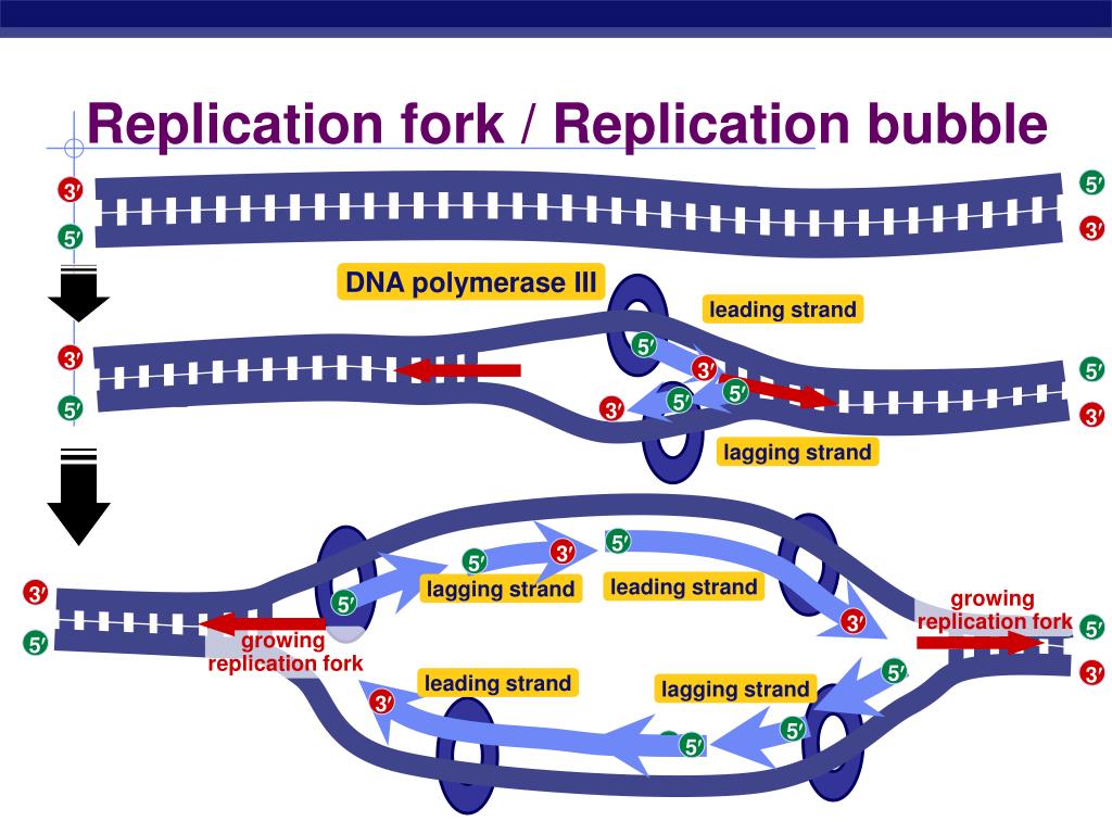
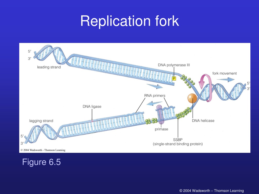
What Happens at the Replication Fork? Two main activities happen at the fork: DNA unwinding and DNA synthesis. The RF unwinds the unreplicated DNA ahead of it through a helicase enzyme.
Replication fork structures acted upon by DNA helicases. A) WRN [72]... Download Scientific
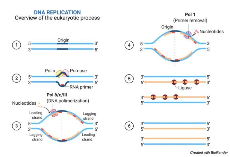
Replication in prokaryotes starts from a sequence found on the chromosome called the origin of replication—the point at which the DNA opens up. Helicase opens up the DNA double helix, resulting in the formation of the replication fork. Single-strand binding proteins bind to the single-stranded DNA near the replication fork to keep the fork open.

Dna replication fork. Biology lessons, Teaching biology, Biomedical science
During replication the two strands of DNA separate; the resulting structure is called the replication fork. The replication fork forms because enzymes called helicases surround the DNA strands and break the hydrogen bonds which hold them together. The result is that two long branches, almost like fork prongs, each of which is a DNA strand.

Dna Ligase Replication Fork Bryce Whitmore
In molecular biology, [1] [2] [3] DNA replication is the biological process of producing two identical replicas of DNA from one original DNA molecule. [4] DNA replication occurs in all living organisms acting as the most essential part of biological inheritance.
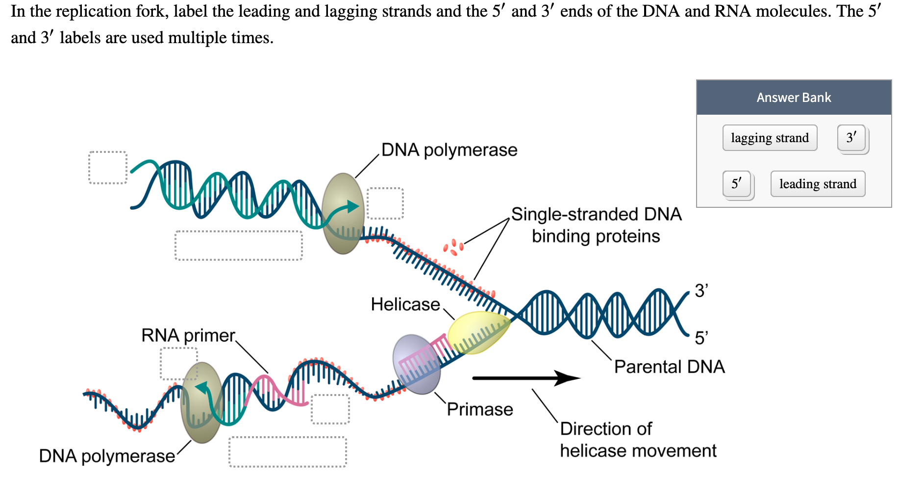
Solved In the replication fork, label the leading and
Two replication forks are formed at the origin of replication and these get extended bi- directionally as replication proceeds.. replication fork Y-shaped structure formed during initiation of replication single-strand binding protein during replication, protein that binds to the single-stranded DNA; this helps in keeping the two strands of.
A, schematic representation of the replication fork. Polymerase III... Download Scientific Diagram
When a cell divides, it is important that each daughter cell receives an identical copy of the DNA. This is accomplished by the process of DNA replication. The replication of DNA occurs during the synthesis phase, or S phase, of the cell cycle, before the cell enters mitosis or meiosis. The elucidation of the structure of the double helix.

DNA Replication Fork Definition & Overview Video & Lesson Transcript
After replication forks are reversed into a 4-way structure and DNA damage is bypassed or repaired, cells need a way to restart DNA replication and restore the replication fork. In addition to using HR ( Figure 2f ), fork restoration can also be accomplished by migrating the reversed fork back into the 3-way junction ( Figure 2e → c ).
[Solved] Draw a schematic diagram of the replication fork, showing Both... Course Hero
In the following diagrams thick lines indicate double stranded duplexes and thin lines indicate individual single strands. A) In our ODIRA model we propose that stalled forks (2a) provide an opportunity for a template switch between the nascent leading strand and the lagging strand template that occurs at short, interrupted inverted repeats (2b).

Replication Fork Diagram Quizlet
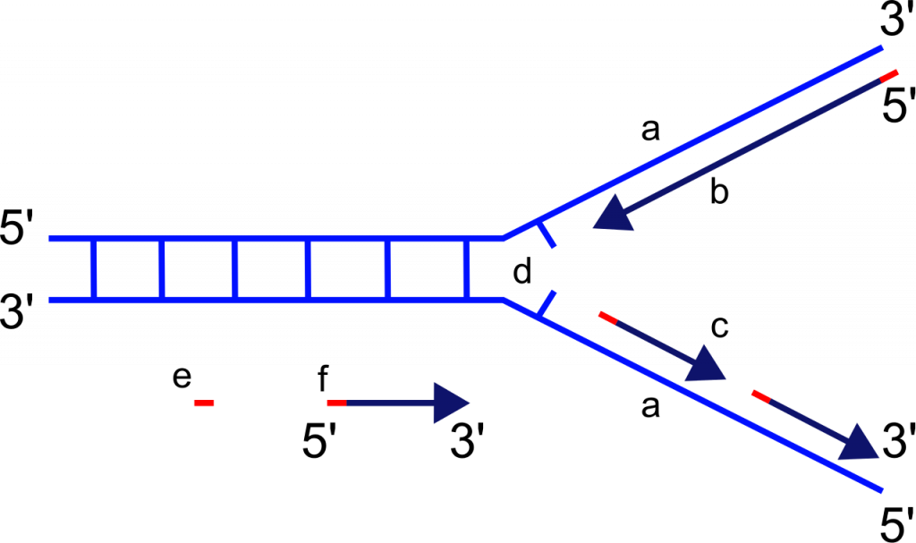
The replication fork is a structure which is formed during the process of DNA replication. It is activated by helicases, which helps in breaking the hydrogen bonds, and holds the two strands of the helix. The resulting structure has two branching's which is known as prongs, where each one is made up of single strand of DNA.
Draw a labelled schematic sketch of replication fork of DNA. Explain the role of enzymes
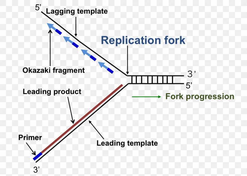
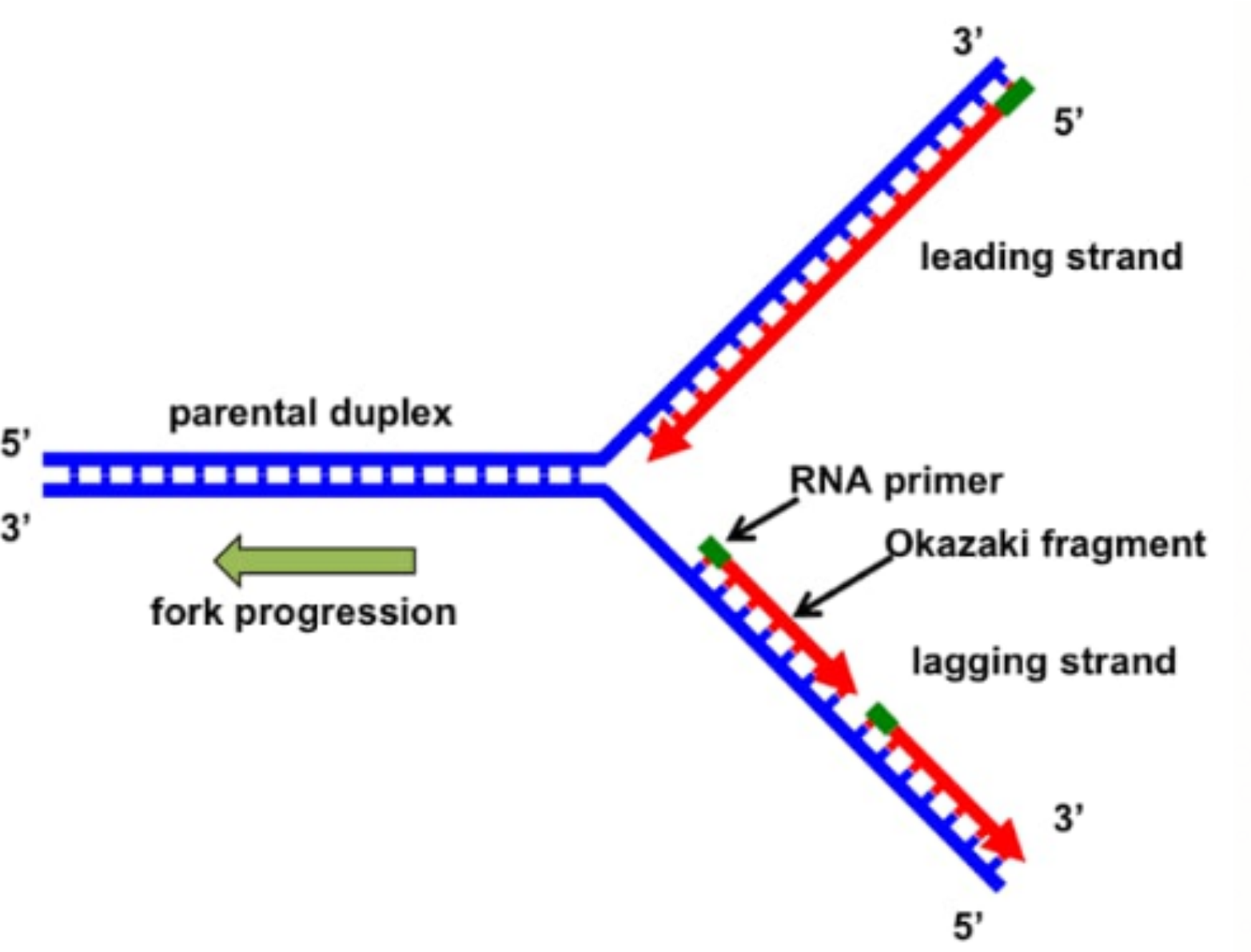
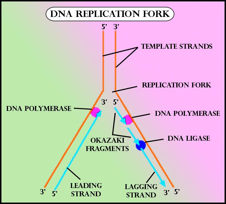
The replication of the other strand, which runs in the 5′ to 3′ direction away from the fork, is made discontinuously. It happens because as the fork moves forward, the DNA polymerase (which is moving away from the fork) comes off and then reattach to the newly exposed DNA. This strand is called the lagging strand.
Draw a labeled diagram of a replicating fork.
Key Terms. origin of replication: a particular sequence in a genome at which replication is initiated; leading strand: the template strand of the DNA double helix that is oriented so that the replication fork moves along it in the 3′ to 5′ direction; lagging strand: the strand of the template DNA double helix that is oriented so that the replication fork moves along it in a 5′ to 3′ manner
PPT DNA Replication PowerPoint Presentation, free download ID2591641
The replication fork * is a region where a cell's DNA * double helix has been unwound and separated to create an area where DNA polymerases and the other enzymes involved can use each strand as a template to synthesize a new double helix. DNA Base RNA Base An enzyme called a helicase * catalyzes strand separation.
PPT Chapter 6 The of PowerPoint Presentation ID9491318
The process of semiconservative replication suggested a geometry for the site of DNA replication, a fork-like DNA structure, where the DNA helix is open, or unwound,. Replication Fork Barriers (RFBs) control DNA progression to protect genomic integrity. RFBs allow for the coordination of DNA replication with important processes on chromatin.
DNA Replication AP® Biology Crash Course Review Albert.io
Step 1: Initiation The point at which the replication begins is known as the Origin of Replication (oriC). Helicase brings about the procedure of strand separation, which leads to the formation of the replication fork. Step 2: Elongation
Replication Fork Definition, Structure, Diagram, & Function
What is the replication fork in DNA? The replication fork is a Y-shaped structure. It forms at the repication bubble with the help of the enzyme DNA helicase. What causes replication fork?.

Simplified model of replication fork Download Scientific Diagram
During DNA replication, both strands of the double helix act as templates for the formation of new DNA molecules. Copying occurs at a localized region called the replication fork, which is a Y shaped structure where new DNA strands are synthesised by a multi-enzyme complex. Here the DNA to be copied enters the complex from the left.